Dermatologists specialize in diagnosing skin cancer using diagnostic tools and expertise. They identify cancer through medical history assessment, dermoscopy, biopsy, and lesion analysis. By detecting early-stage cancers, you can access comprehensive treatment options and better long-term outcomes. Here is how dermatologists diagnose cancer:
Reviewing Patient’s Medical History
Your medical history can provide doctors insight when identifying cancers. Dermatologists ask about family cancer history, past sun exposure, suspicious lesions, and lifestyle habits. They inquire about prior cancers, sunburns, suspicious moles, smoking, and occupational chemical exposure. Clinical context provides clues for identifying hereditary or lifestyle risks that may impact cancer risk and outcomes. Providing your past medical records and longitudinal data helps doctors recognize disease patterns. This allows them to create a diagnostic approach tailored to you.
Using Dermoscopy Technology
Dermoscopy technology uses polarized light and magnification to reveal subsurface pigmentation, vascular patterns, and structural details. This helps differentiate harmless moles from early melanomas or basal cell carcinomas needing a biopsy. Some dermatoscopes use ultraviolet imaging to make changes invisible to the naked eye visible.
Dermatologists analyze the results of this procedure to identify unusual textures, blood vessels, colors, and lesions that may need further tests. The enhanced imaging can give doctors a more precise picture that helps determine the next steps. Dermatologists may use total body photography to track changes in moles and lesions over time in high-risk patients. This method involves taking detailed photographs of the entire skin surface to establish a baseline for future comparisons.
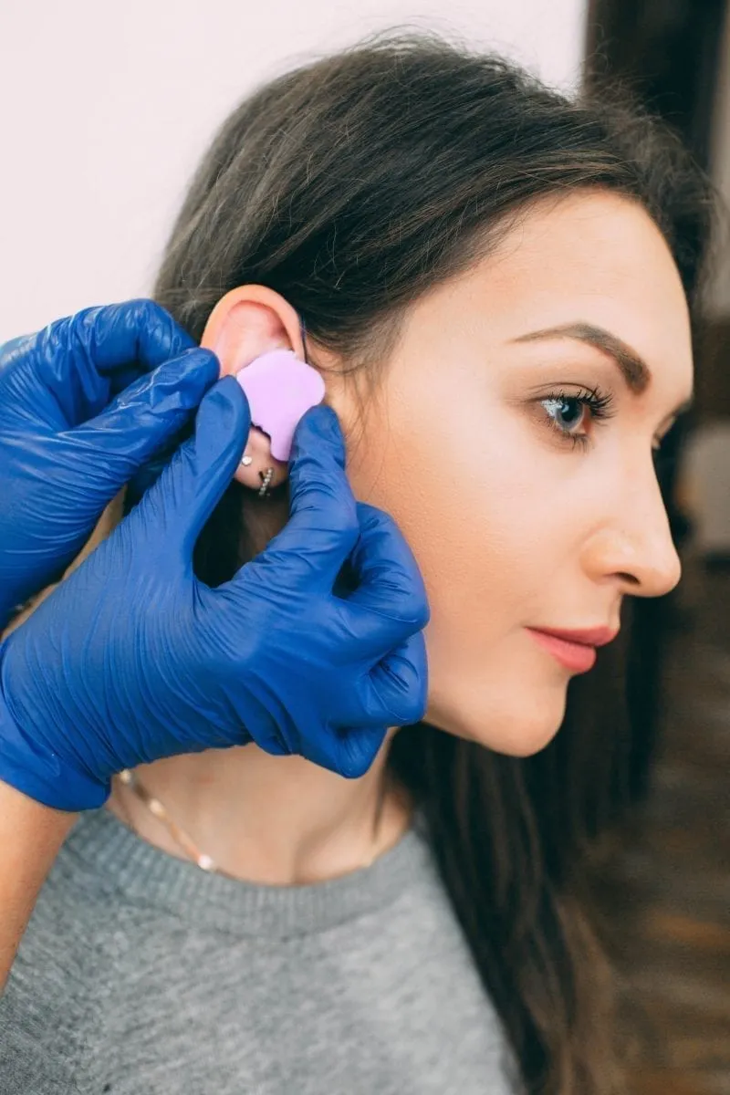
Performing Biopsy Procedures
Suspected cancer lesions identified through visual and dermoscopic examination require biopsies. Dermatologists use excisional, punch, shave, and incisional biopsies to analyze abnormal cells. Pathological examination of biopsy samples identifies cancer cell types or confirms cancer. This confirms diagnosis accuracy and provides information on how advanced the cancer is. You can receive personalized treatments based on your cancer’s attributes and characteristics.
Analyzing Skin Lesions
Dermatologists diagnose skin lesions by evaluating their diameter, borders, color, symmetry, and surface textures. Cancerous lesions possess assorted pigmentations, uneven borders, and rapid vertical spread. If an existing mole changes size, shape, or color, that often signals malignancy. Dermatologists monitor lesions meeting any criteria for diagnostic certainty. Early detection of skin cancer offers more treatment opportunities that improve patient outcomes, including supporting cancer treatments and cancer prevention.
Identifying Early Symptoms
Dermatologists are trained to recognize the early symptoms of cancer, including sores that do not heal, scaly patches of skin, or ulcers. Detecting symptoms early helps prevent cancer from progressing to more severe stages. Regular skin checks enable tracking of any changing or newly emerging lesions. Early-stage detection of lesions enables dermatologists to remove malignant cells before they begin spreading. Dermatologists then focus on preventing recurrence and on the early treatment of cancer.
Test for Skin Cancer Today
Dermatologists diagnose skin cancer using their expertise and specialized tools to detect abnormal lesions early and inform treatment. It often takes two to three weeks to get the results of a biopsy, and if tests are positive, doctors may recommend chemotherapy creams to treat the area. If you suspect you have skin cancer, schedule a consultation today.